Q:
How is osteoporosis diagnosed?
A:
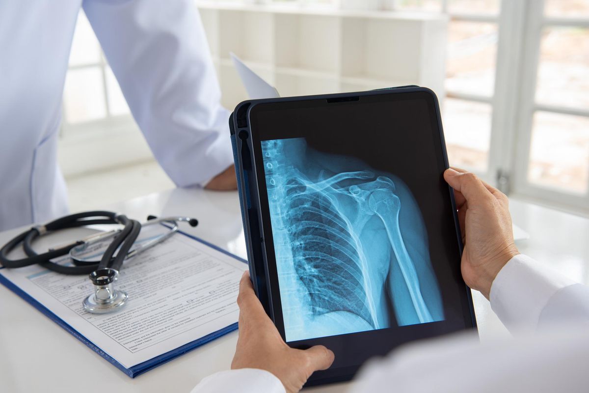
Osteoporosis develops gradually, usually without symptoms. A broken bone that occurs with minor trauma, such as a fall onto an outstretched hand that breaks the wrist, is typically the first symptom. Your health care professional can make a diagnosis of osteoporosis based on your medical history with an assessment of your risk factors; a physical examination and laboratory tests; and a bone mineral density (BMD) test, a noninvasive test that measures your bone mass.
Other common risk factors for osteoporosis are: age—your risk for fractures increases as you go from your 50s to 60s to 70s and older; small, thin frame (weighing less than 127 pounds); personal and/or family history of broken bones as people age, especially a history of spine or hip fractures in an elderly parent; previous history of fractures after age 45; low lifetime intake of calcium; excessive thinness; smoking; excessive alcohol consumption; inactive lifestyle; estrogen deficiency caused by menopause and certain medical conditions such as anorexia nervosa; prolonged absence of menstrual periods as a young woman; long-term use of corticosteroids and some anticonvulsants; Caucasian or Asian ethnic backgrounds, but older African-Americans and Hispanic Americans are also at significant risk; certain chronic medical conditions including diabetes, hyperthyroidism, hyperparathyroidism, rheumatoid arthritis and some bowel diseases that cause poor absorption of calcium or vitamin D; depression; and long-term use of proton pump inhibitors.
Your health care professional may also recommend laboratory tests to help identify or rule out conditions other than menopause that may be causing low bone density. These tests include a complete blood cell count and serum chemistry studies. If your medical history or test results suggest causes of bone loss other than menopause and age, then additional laboratory tests may be conducted.
The U.S. Preventive Services Task Force (USPSTF) recommends that women age 65 and older without previous fractures or other risk factors be routinely screened for osteoporosis using a bone density test. The USPSTF also recommends screening for women younger than age 65 who have a fracture risk equal to or greater than that of the typical 65-year-old woman. In addition, the National Osteoporosis Foundation and the International Society for Clinical Densitometry recommend screening in postmenopausal women and men 50 to 70 who have risk factors for the disease. The National Bone Health Alliance recently adopted new criteria that call for diagnosing osteoporosis in anyone over age 50 who has demonstrated an increased risk for fractures.
Talk to your health care practitioner about when you should be screened. And have your BMD screening performed at a physician's office, hospital or osteoporosis center that does bone density testing regularly.
There are several types of BMD tests, including the following:
- DEXA (dual energy X-ray absorptiometry) measures bone density at the spine, hip or total body. This is the standard test for making a diagnosis.
- QCT (quantitative computed tomography) may be used to measure the spine as an alternative to DEXA. This test is rarely used because it is expensive and requires a higher dose of radiation.
- Ultrasound uses sound waves to measure bone density, and it may be used to measure bone density at the heel, which may be useful in determining a person's fracture risk. Ultrasound is used less often than DEXA because there are no guidelines that use ultrasound measurements to predict fracture risk or diagnose osteoporosis.
Results of BMD tests done on postmenopausal women and older men are usually expressed as "T-scores," a measure of how far your bone density deviates above or below the average bone density value for a young, healthy, Caucasian woman.
- A T-score between +1 and -1 indicates normal bone density.
- A T-score between -1 and -2.5 usually signals low bone density.
- A T-score at or below -2.5 usually signals osteoporosis.
Your bone density is also compared to an "age matched" standard. The age-matched reading (Z-score) compares your bone density to the "norm" for your age, sex and size. You may be normal for your age and still have significant low bone mass or osteoporosis. The T-score is more important for women over 60.
Your T-score will help your health care professional determine whether you are at risk for a fracture. Generally, the lower your bone density, the higher your risk for fracture. However, your health care professional will consider your BMD score along with your age, personal health history, osteoporosis risks and lifestyle, including whether you exercise and are getting adequate calcium and vitamin D.
Your health care professional may also use a fracture risk assessment tool (FRAX). Your FRAX score, which is based on your age, weight, height and presence or absence of several important risk factors, as well as your bone mineral density at the hip, determines your risk of having a hip fracture and several other types of osteoporotic fractures in the next 10 years. This information can be used to determine if medical treatment is necessary to lower your risk of serious fractures.
By weighing all of these factors, your health care professional can determine if osteoporosis poses a significant threat for you now or in the years ahead.
Some tests for osteoporosis risk, such as those available at community health fairs, provide a starting point for assessing your bone health—but definitely require follow-up. If you have one of these types of tests, be sure to discuss the results with your health care professional, especially if your results indicate low bone density.
To learn more about osteoporosis and preventing broken bones, visit
https://healthywomen.org/condition/osteoporosis.
- Covid-19 Poses Challenges for Osteoporosis Treatment and Diagnosis - HealthyWomen ›
- My Family’s History With Osteoporosis Changed My Medical Career Path - HealthyWomen ›
- Taking on Mountains — and Osteoporosis - HealthyWomen ›
- Taking on Mountains — and Osteoporosis - HealthyWomen ›
- How Do Hormones Affect Osteoporosis? - HealthyWomen ›
- What Increases Your Risk for Bone Fractures and Osteoporosis? - HealthyWomen ›
- Osteoporosis and Fractures: What You Should Know - HealthyWomen ›
- Osteoporosis Treatment - HealthyWomen ›
- Osteoporosis Is a Looming Public Health Crisis - HealthyWomen ›


